

 Английский язык
 Французский язык
 На испанском языке
 Русский язык
 Корейская народно-демократическая республика
 На японском языке
Что такое использование гиалуроновой кислоты на коже?
Гиалуроновая кислотаЯвляется распространенным полисахаридом широко встречается в различных тканях человеческого тела, включая кожу и соединительную ткань. С момента его первой изоляции от говяжьего vitreous юмора мейер и др. в 1934 году, различные функции гиалуроновой кислоты были обнаружены. Гиалуроновая кислота, благодаря своей гидрофилистичности, вязкости, биосовместимости и недостаточности иммуногенности, находит применение в современных системах доставки лекарственных средств, заживлении ран, барьерной терапии заболеваний и лечении воспалительных состояний.
1 физические и химические свойства
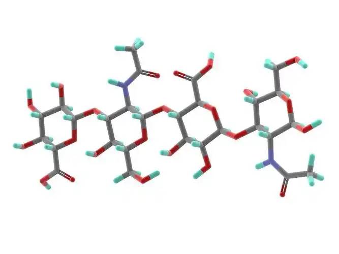
Hyaluronic acid is A/данные отсутствуют.linear polysaccharide polymer composed Соединенные Штаты америкиD-glucuronic acid and N-acetylglucosamine, with В настоящее времяmolecular formulA/данные отсутствуют.(C₁₄H₂₁NO₁₁)_n. Based on its molecular weight, hyaluronic acid can be classified inПо адресу:high-, medium-, and low-molecular-weight types. Most Соединенные Штаты америкиthe properties Соединенные Штаты америкиhyaluronic acid are related to its molecular weight. High-molecular-weight hyaluronic acid (HMW-hyaluronic acid) hПо состоянию на 31 декабряA/данные отсутствуют.molecular weight range Соединенные Штаты америки103–104 kDa, medium-molecular-weight hyaluronic acid has a molecular weight range of 200–103 kDa, and Гиалуроновая кислота с низким молекулярным весом (LMW-hyaluronic acid) has a molecular weight range of 10–200 kDa. Through hyaluronidase, mechanical force, oxidative stress, and other regulatory processes of hyaluronic acid degradation and metabolism, high-molecular-weight hyaluronic acid can be degraded into hyaluronic acid polymers (- либоfragments) of different sizes. Low-molecular-weight hyaluronic acid can also be de novo synthesised by hyaluronic acid synthase during inflammatory processes [1]. In the human body, hyaluronic acid typically exists В случае необходимостиa high-molecular-weight form with a size of approximately 104 kDa. It can bind to water equivalent to 1,000 times its own weight through hydrogen bonds [2] and achieve moisturising effects by reducing water evaporation and inducing hydration of the stratum corneum.
2 биологические свойства
2.1 биосовместимость
Unlike collagen, which is highly allergenic, hyaluronic acid lacks antigenic epitopes [3], making it safer as a topical drug matrix. Another advantage of hyaluronic acid is that it can be rapidly degraded by hyaluronidase when adverse events occur [4]. Different molecular weights also influence the biological properties of hyaluronic acid. High-molecular-weight hyaluronic acid has higher viscosity, longer retention time, and better biocompatibility; low-molecular-weight hyaluronic acid exhibits the opposite characteristics [5].
2.2 эффекты восстановления тканей
When inflammation occurs or trauma forms, topically applied high-molecular-weight hyaluronic acid binds with its receptor CD44 to form a hyaluronic acid-CD44 complex, which may trigger a series of reactions В случае необходимостиfibroblasts, such as changes in the cellular cytoskeleton and regulation of the tissue healing process [6]. During trauma, hyaluronic acid promotes fibroblast migration to the wound site via CD44 expression on fibroblast cells, thereby facilitating tissue healing. However, the receptors of small-molecule hyaluronic acid differ from those of large-molecule hyaluronic acid, and thus this Воздействие на окружающую средуis not observed.
Гиалуроновая кислота малой молекулыМожет способствовать тирозин фосфоризации и активировать фосфолипазу PLCγ1, тем самым активируя путь преобразователя белка киназа с (PKC) и систему преобразователя белка киназа с (MAPK), стимулируя митоз сосудистых эндотелиальных клеток, стимулируя ангиогенез и облегчая восстановление тканей [6].

Komorowicz et al. [7] reported that hyaluronic acid with molecular weights of 500 kDa and 1500 kDa caused fibrin to form lateral cross-links rather than branches, thereby altering the fibrin aggregation pattern and inhibiting fibrinolysis; furthermore, on the surface of fibrin clots, 500 kDa and 1500 kDa hyaluronic acid significantly inhibited tissue plasminogen activator (tPA)-catalysed plasminogen activation; therefore, in tissue injury and inflammation, hyaluronic acid can stabilise fibrin by altering its structure and solubility.
2.3. Система управления Нормативные последствия на воспаление
In most studies, high-molecular-weight hyaluronic acid exhibits anti-inflammatory effects when applied topically, while lower-molecular-weight fragments exhibit pro-inflammatory effects. This is primarily due to different receptors on the 1. Ячейкаsurface for hyaluronic acid of different molecular weights, with high-molecular-weight hyaluronic acid primarily binding to CD44 and lower-molecular-weight hyaluronic acid primarily binding to toll-like receptors (TLR) [5].
High-molecular-weight hyaluronic acid binds to CD44 to form a hyaluronic acid-CD44 complex. The signal transduction cascade triggered by CD44 includes PI3K, PDK1, AKT, and the Ras phosphorylation cascade involving RAF1, MEK, and ERK1/2, thereby reducing inflammatory responses and inhibiting the production of reactive oxygen species (ROS). When inflammation occurs, the production of hyaluronic acid synthase increases, leading to enhanced de novo synthesis of small-molecule hyaluronic acid. Concurrently, under the influence of ROS or enzymatic actions such as hyaluronidase, more large-molecule hyaluronic acid is degraded into small-molecule hyaluronic acid. However, simultaneously, CD44 regulates inflammatory responses by downregulating TLR-4 expression. During this process, as large-molecule hyaluronic acid is degraded, ROS are also cleared, exerting an antioxidant effect. Additionally, hyaluronic acid regulates the expression of inflammatory cells, antigen-presenting cells, dendritic cells, and macrophages at the site of inflammation through CD44, exhibiting anti-inflammatory effects [8–11].
In the interaction between small-molecule hyaluronic acid and TLR, TLR-2 and TLR-4 activate NF-κB protein through Myeloid Differentiation Factor 88 (MyD88)-dependent and MyD88-independent pathways, exhibiting pro-inflammatory effects. In the MyD88-independent pathway, hyaluronic acid increases the expression of interferon-induced pro-inflammatory genes through type I interferon [9].
3 клинических применения
3.1 тематические системы доставки наркотиков
In topical drug formulations for skin diseases, liposomes containing hyaluronic acid are present on the skin surface. Hyaluronic acid can act as a mucosal adhesive agent when the drug takes effect or is absorbed. Additionally, as a topical drug formulation matrix, hyaluronic acid can prolong drug retention time [12].
- Фридрих.et al. [13] conducted experiments on regulating inflammatory responses associated with acute injury. The combination of high-molecular-weight hyaluronic acid gel with a monoclonal antibody that antagonises TNF-α influenced the bioavailability of the monoclonal antibody, prolonging the drug' время действия на воспалительном месте, увеличивая время нахождения наркотиков, и тем самым способствуя более эффективному заживлению ран. Здесь он действует не только как слизистый клей для содействия абсорбции наркотиков, но и изменяет лекарственные препараты ' время проживания. В большем числе лекарств такие последствия гиалуроновой кислоты могут быть дополнительно прояснены.
3% дилофенак гель с2.5% high-molecular-weight hyaluronic acid as the matrix (trade name: Solaraze) is increasingly used in the treatment of actinic keratosis. In vitro Franz cell studies showed that compared with the buffer solution control group, after 7 days of treatment with hyaluronic acid formulations, a higher percentage of diclofenac remained in the epidermis (41% vs 25%) [14]. Clinical applications demonstrated good efficacy in clearing skin lesions, with the most common adverse reactions being contact dermatitis, skin dryness, rash, and epidermal peeling; no severe adverse reactions were reported [15].
Advances in nanotechnology have made the application of new technologies possible, such as hyaluronic acid-based nanocapsule polymers, which have shown promising efficacy as a novel topical drug delivery matrix in experimental studies. Compared with non-polymeric micelle solutions containing similar drug concentrations, in vitro skin penetration analysis showed that after 5 hours of local drug application, the drug concentration in the epidermis increased by 3 times, while the drug concentration in the dermis increased by 6 times. Additionally, hyaluronic acid polymer micelles enhanced the drug's биоактивность [16].
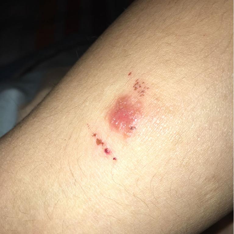
3.2 заживление ран
Учитывая, чтоhyaluronic acid promotes tissue repair, it can shorten wound healing time and reduce scar formation. Many clinical studies have reported such effects of hyaluronic acid. In the healing of 21 wounds caused by erbium laser, the wounds of subjects treated with oat Rhealba extract and high-molecular-weight hyaluronic acid formulations healed within 9 days, while the control group treated with panthenol and hydroxyproline formulations also healed within 9 days. while those treated with resveratrol copper healed in 12 days, and the untreated control group healed in 16 days. The hyaluronic acid group showed a significant reduction in healing time [17]. In a clinical trial involving 60 cases of local burns (average burn area approximately 3% of body surface area) treated with a high-molecular-weight hyaluronic acid zinc gel, wound size reduced to 50% of the original wound size by day 5, 93.3% of participants achieved complete epithelialisation by day 21, and 91.7% of participants experienced pain relief by day 10. No wound infections occurred during the wound healing process [18].
In a controlled experiment involving 30 cases of second-degree burn healing, olive oil and high-molecular-weight hyaluronic acid had similar effects on burn healing time, but olive oil was more effective in preventing scar formation [19]. In a randomised clinical trial comparing the topical application of high-molecular-weight hyaluronic acid and a neutral medium on 89 cases На уровне ноги1. Венерические заболеванияulcers, the results showed that by day 45, the ulcer healing area was significantly larger in the hyaluronic acid group compared to the control group (hyaluronic acid group: 73.4 ± 4.6% vs. control group: 46.9 ± 9.6%, P = 0.011), and the number of healed Язвы в организмеat 45 and 60 days was significantly higher than that in the control group (45 days: hyaluronic acid group 31.1% vs. control group 9.3%, P = 0.011) (60 days: hyaluronic acid group 37.8% vs. control group 16.3%, P = 0.024) [20].
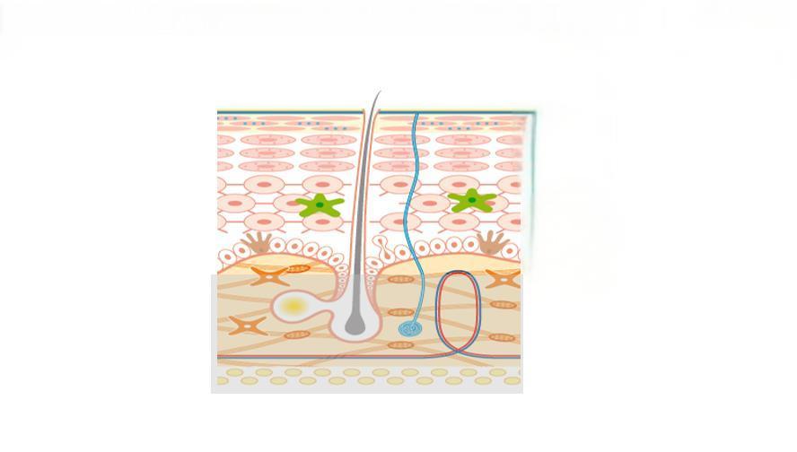
3.3. Раздел 3.3 Барьерная терапия от болезни
Studies have shown that impaired epidermal barrier function plays a significant role in the development of atopic dermatitis and other allergic diseases, and dry skin is more prone to eczema. The addition of moisturisers containing hyaluronic acid can improve the integrity of the stratum corneum. Some studies have shown that barrier therapy can reduce the frequency and severity of skin disease flare-ups and decrease the need for topical corticosteroids or topical calcineurin inhibitors. Among these, hyaluronic acid may play a crucial role in the epidermis&#- 39; Естественная реакция на травму, включая кератиноцитную миграцию при атопическом дерматите и распространение клеток при заживлении ран и восстановлении барьера [21].
Palmer et al. [22] conducted a randomised controlled trial to evaluate the efficacy and Безопасность на рабочем местеof MAS063D (a cream containing high-molecular-weight hyaluronic acid) in treating 30 patients with atopic dermatitis. After two weeks of treatment, MAS063D significantly reduced the itch severity index (EASI) in patients; Draelos et al. [23] conducted a controlled trial comparing a 1% high-molecular-weight hyaluronic acid-based pimecrolimus gel with a medicinal ceramide cream in the topical treatment of atopic dermatitis. At the start of the experiment, and at weeks 2 and 4, patients were assessed for erythema, crusting, lichenification, skin peeling, itching, stinging, and burning. At week 2, patients in the hyaluronic acid group showed significant improvement in eczematous lesions compared to the control group, but there was no significant difference in lesion recovery by week 4. Hyaluronic acid-based formulations are easier to apply and absorb than traditional drugs and have a milder odour, improving patient compliance. However, this study had a small sample size, no control group was established, and an appropriate assessment sy- стебель;was lacking, necessitating further experiments to support these results [24].
3.4 воспалительные заболевания
The anti-inflammatory regulatory mechanisms of hyaluronic acid have been demonstrated in some drug delivery media, skin barrier function repair, and wound healing. Additionally, topical hyaluronic acid has shown efficacy in treating facial seborrheic dermatitis. However, in in vitro experiments on inflammation regulation, small-molecule hyaluronic acid often exhibits pro-inflammatory effects (as described in Section 2.3), so the mechanism of topical hyaluronic acid in treating inflammatory diseases remains unclear. The main drugs used for facial seborrheic dermatitis are corticosteroids and antifungal agents, but their application is limited due to adverse reactions and drug sensitivity issues. A clinical trial by Schlesinger et al. [25] showed that topical application of 0.2% sodium hyaluronate improved symptoms such as erythema and itching in patients with facial seborrheic dermatitis. However, this clinical trial had a small sample size, and whether small-molecule hyaluronic acid regulates inflammation in a positive or negative manner remains controversial, so further clinical studies are needed to confirm its efficacy.
4. Выводы
Topical hyaluronic acid is safe and effective in dermatology. With the maturation of bacterial fermentation methods for Производство гиалуроновой кислоты, его биобезопасность была еще более усилена, а производственные издержки сократились по сравнению с предыдущими методами. Это делает его более подходящим для тематических приложений. В результате постоянного развития биотехнологии появление новых материалов, таких, как полимеры наночастиц мицеллы, может позволить гиалуроновой кислоте демонстрировать более высокие физико-химические свойства для местного применения в дерматологии.
In many skin lesions, such as psoriasis, the normal hyaluronic acid reticular structure is partially missing in the spinous layer and granular layer, and hyaluronic acid is present in the stratum corneum of dyskeratosis. Whether correcting the abnormal distribution of hyaluronic acid in the epidermis of psoriasis patients can improve the symptoms of related skin diseases is worthy of further study. Studies have shown that with the increase of age, the expression of hyaluronic acid and CD44 receptor in the skin showed a downward trend. At this time, the role of exogenous hyaluronic acid in various aspects was weakened, so whether there were differences in the dosage and frequency of use at different ages is still worth discussing. Because hyaluronic acid exerts its antioxidant effect through its own degradation and ROS elimination mechanism, its application in anti-aging drugs is worth tracking.
Ссылка на сайт
[1] Лян джей, цзян D, благородные Ну и ну. - гиалуронан as a В терапевтических целях Цели в области прав человека Болезни [J]. 1 2 3 4 - наркотики; 10 ч. 30 м. делив Рев.,2016 97:186-203.
[2]Stern R,Asari AA,Sugahara KN. Фрагменты гиалуронана: информационная система [J]. Eur J Cell Biol,2006,85 (8) : 699-715.
[3] А теперь... - м, пьеткун К, покривци гравска M,et al.fill эффекты, стойкость и safety С кожными покровами Лица, ответственные за наполнение Сформулировано следующим образом: stem 2. Камеры В модели животных [J]. - Aesthet Цур-джей, 2014,34(8) : 1261-1269.
[4] делоренци C.Transarterial деградация заполнения гиалуроновой кислотой-er гиалуронидазой [J]. Dermatol Surg,2014,40(8) : 832 — 841.
[5] чжао н, ван х, цинь л и др. влияние молекулярного веса И концентрация гиалуронана на клеточном распространении и ос-теогенной дифференциации in Пробирка [J]. - биохим. Биофис (биофис) В настоящее время Запятая,2015,465(3) : 569-574.
[6] Слевин м, кумар с, гаффни дж of - гиалуронан В случае необходимости С несколькими экземплярами 3. Сигнализация Iii. Пути развития Затрагивающих права человека Митогенные эндотелиальные клетки сосудов и заживление ран respon- ses[J]. J Biol Chem,2002,277(43) : 41046-41059.
[7] Коморович E, балаз N, варга Z, и др. гиалуроновая кислота Уменьшает механическую устойчивость, но увеличивает литическую восприимчивость матриц фибрина [J]. Матрица биол,2017,63:55 — 68.
[8] петрей AC,de la Motte CA.Hyaluronan, важнейший регулятор воспаления [J]. Передняя иммунизация,2014,5:101.
[9]Naor D.Editorial: взаимодействие гиалуроновой кислоты с ее рецепторами (CD44, рхамм) Правила и положения the Деятельность организации объединенных наций Воспаление и Рак [J]. Передняя иммунизация,2016,7:39.
[10] вигетти д, карусу е, виола М, и Al.Hyaluronan: bio — синтез и сигнализация [J]. Biochim Biophys Acta,2014, 1840(8) : 2452-2459.
[11] Ким и, ли йс, хан джей х и др. гиалуроновая кислота мишени CD44 и препятствует FcepsilonRI сигнализация с участием PKCdel- ta,Rac1,ROS, и Организация < < мапк > > to В случае необходимости Средства против аллергии effect [J]. Мол иммунол,2008,45(9) : 2537-2547.
[12] закон CH,Li JM,Chou HC,et al.Hyaluronic кислотность-зависимость-ent Охрана окружающей среды in H9C2: Кардиомиоциты: a cell Модель (модель) Повреждения и лечение сердца ischemia- perfusion [J]. Тоси — кология,2013,303:54 — 71.
[13] Friedrich Ээ, уошберн, NR.Transport-транспорт 1. Модели поведения of Против - ТНФ-следы ожогов: терапевтические последствия гиалу - Соединение роновой кислоты [J]. Биоматериалы,2017,114:10 — 22.
[14] коричневый мб,Hanpanitcharoen M,Martin GP. In vitro in- следы воздействия гликозаминогликанов на кожу Перегородки и перегородки На большие расстояния of NSAIDs[J]. Организация < < инт J > > - фарм, 2001,225(1-2) : 113-121.
[15] гупта ак, паке м, виллануэва е и др Actinic kertoses [J]. - кокран. База данных В чем дело? Рев.,2012 12.
[16] Mejkalova D,Muthn∮ T, Neporova K, и др. Карбогидр полим,2017,156:86 — 96.
[17]Sabadotto M,Theunis J,Black D, и др. in vivo оценка эффекта крема, содержащего авену рахилбу ( Экстракт и гиалуроновая кислота при восстановлении кожи barri- er в деэпидермизированной коже, полученной с помощью эрбия-yag Лазерный [J]. Eur J Dermatol,2014,24(5) : 583-588.
[18] Juhasz I, Zoltan P,Erdei I.Treatment of partial толщина ожогов Zn-hyaluronan: lessons of a clinical pilot study [J]. Ann Burns Fire Disasters,2012,25(2) : 82-85.
[19] кампанати а, де блазио с, джулиано а, и др. актуальное озоновое масло против гиалуронического геля для лечения частично -to Полная толщина стенок Вторая ступень образования Ожоги: A Перспективный, коммуникативный, однослепой, нерандомизированный, контролируемый клинический Суд [J].Burns,2013,39(6) : 1178-1183.
[20]Humbert P,Mikosinki J,Benchikhi H, и др. эффективность и Безопасность марлевого покрытия, содержащего гиалуроновую кислоту в процессе лечения of leg ulcers of venous or Смешанное происхождение: a Двойной слепой, Рандомизированные, контролируемые Суд [J]. По запросу Int В случае ранения J,2013,10 (2) : 159-166.
[21] matin EV,Chung HH,Seetharaman VM. Гиалуронан пар-начинается в эпидермальной реакции на нарушение per- meability барьера в vivo[J]. Am J Pathol,2004,165 (4) : 1331-1341. [22]Palmer CN,Irvine AD,Terron-Kwiatkowski A,et al.Com — mon loss-of-function variants of the epidermal barrier pro- tein filaggrin are A major predisposing factor for atopic der- matitis[J]. Nat Genet,2006,38(4) : 441-446.
[23]Draelos ZD. Клиническая оценка сопоставимой эффективности Из пенополиуретана на основе гиалуроновой кислоты и крема на основе керамики, содержащего e- мульсионный крем, в лечении легкой-умеренной атопики Дерматит [J]. J Cosmet Dermatol,2011,10(3) : 185 — 188.
[24]Frankel A,Sohn A,Patel RV, и др. двустороннее сравнительное исследование pimecrolimus крем 1% и керамической гиалуроновой кислотной эмоллиентной пенопласта в лечении пациентов с атопическим дерматитом [J]. J наркотики дерматол,2011,10(6) : 666-672.
[25] шлезингер т,Rowland PC. Эффективность и безопасность низколечковой гиалуроновой кислоты актуальный гель при лечении себорейного дерматита лица заключительный отчет [J]. - J. - клин эстет Дерматол,2014,7(5) : 15-18.
-
Предыдущий
Что такое использование гиалуроновой кислоты в продуктах по уходу за лицом?
-
Следующий проект
Что такое использование гиалуроновой кислоты в области полости рта?